PSMA PET-CT scans are important in the detection and staging of prostate cancer. In Australia, the most commonly used tracer for PSMA PET-CT scans is Gallium-68 (Ga-68).
However, Qscan Group uses Fluorine-18 (F-18), a tracer developed by Professor Martin Pomper, director of nuclear medicine and imaging at the Johns Hopkins University School of Medicine.
Benefits of F-18
Improved image quality compared to scans using Ga-68
Intrinsic physical properties, which lends an improved spatial resolution of 5mm
Longer half-life, meaning increased reliability as a tracer
Lower urinary excretion
Improves detection of recurrences and additional lesions
Improved specificity in staging
Higher tumour uptake
High sensitivity, even in small tumours
Improved confidence for EPE and SVI
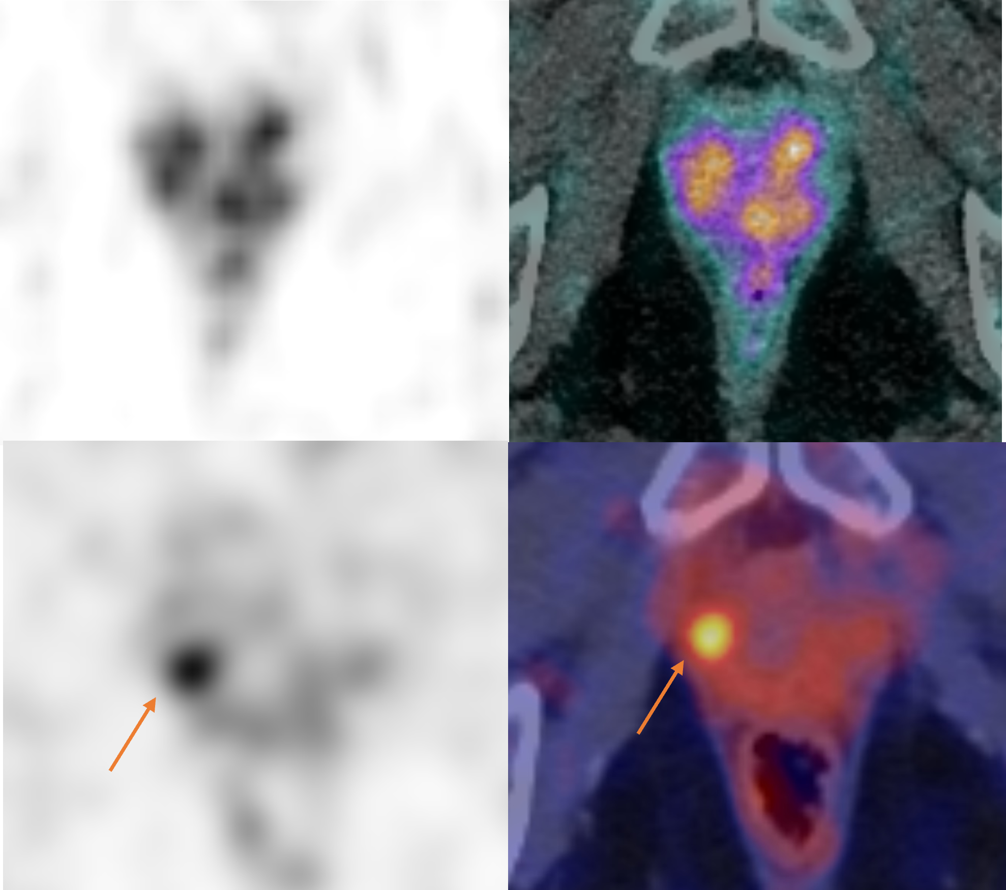
An 83-year-old male with a large basal PZ tumour. SVI is not clear in the Ga-68 PSMA images (note resolution and background noise), but with F-18, SVI is clearly resolved.
Reduced false positives for M staging
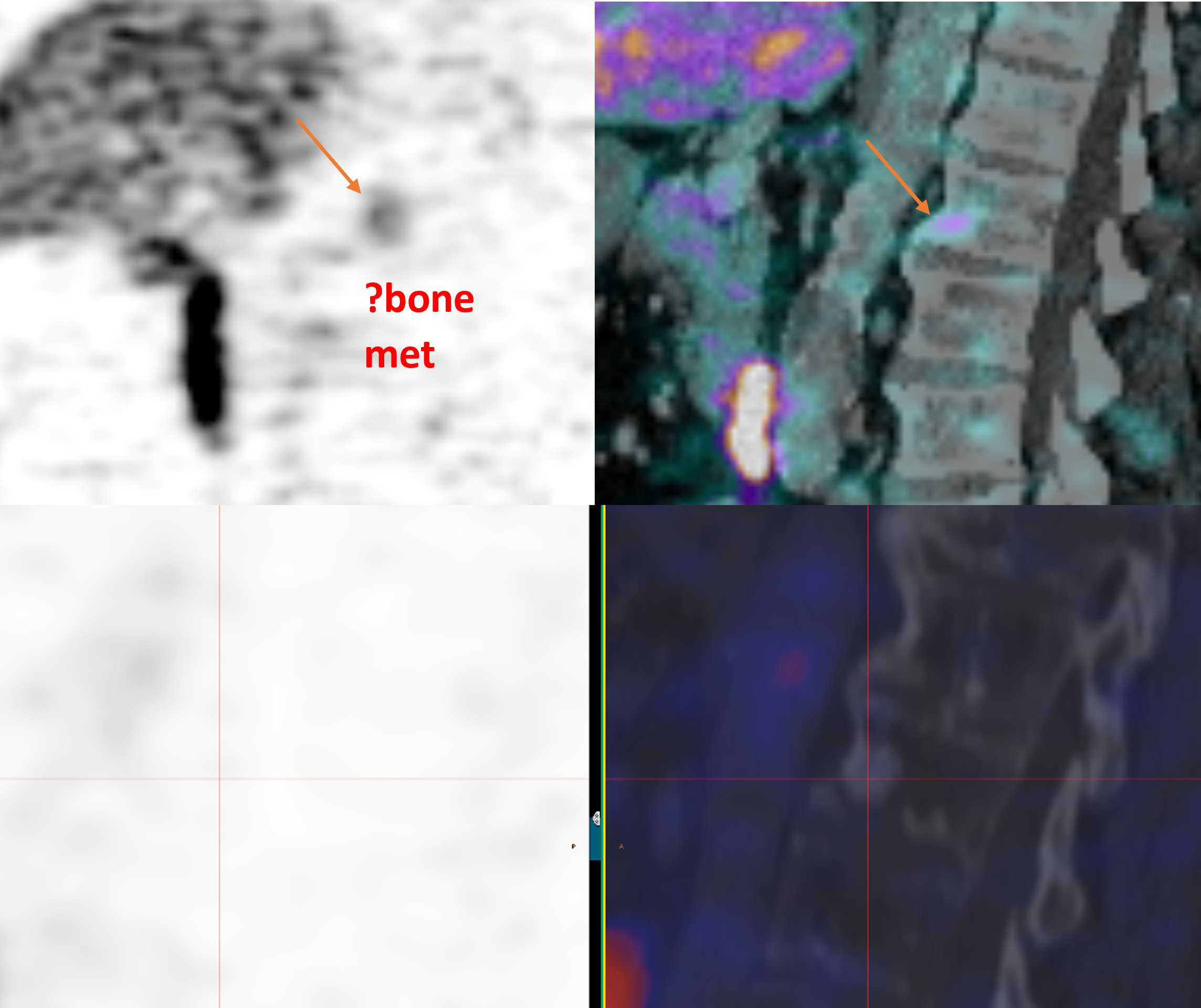
Scans of a 67-year-old male showing uptake in an L2 vertebral sclerotic lesion. Ga-68 PSMA (left) only showed background noise, while F-18 PSMA (right) allowed the patient to be cleared of metastatic disease.
Qscan Group has been using F-18 as our PSMA tracer of choice since 2017.
We are the most experienced in Australia when it comes to using F-18 for PSMA imaging, and will bring this leading expertise to all your prostate patients.
·Brauchli D, Singh D, Chabert C, Somasundaram A, Collie L. Tumour-capsule interface measured on 18F-DCFPyL PSMA positron emission tomography/CT imaging comparable to multi-parametric MRI in predicting extra-prostatic extension of prostate cancer at initial staging. J Med Imaging Radiat Oncol. 2020 Dec;64(6):829-838.
·Parathithasan N, Perry E, Taubman K, Hergarty J, Talway A, Wong L, Sutherland T. Combination of MRI prostate and 18F-DCFPyl PSMA PET/CT detects all clinically significant prostate cancers in treatment-naive patients: An international multicentre retrospective study. J Med Imaging Radiat Oncol 2022 Feb